Back Of Skull Anatomy : Medicine Anatomy Skeleton Bones Skull Back View Steel Stock Photo Alamy - Learn about anatomy skull with free interactive flashcards.
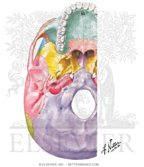
Back Of Skull Anatomy : Medicine Anatomy Skeleton Bones Skull Back View Steel Stock Photo Alamy - Learn about anatomy skull with free interactive flashcards.. The base of the skull (or skull base) forms the floor of the cranial cavity and separates the brain from the structures of the neck and face. Anatomical structures of the skull include: The bbc is not responsible for the content of external websites. Anatomical structures of the skull include: Human anatomy for muscle, reproductive, and skeleton.
A thorough description is beyond the. The skull has a single occipital condyle.7 the skull consists of five major bones: The bbc is not responsible for the content of external websites. The skull begins to form prior to week 12 of embryogenesis. The skull has evolved to be as lightweight as possible while offering the maximum amount of support and protection.
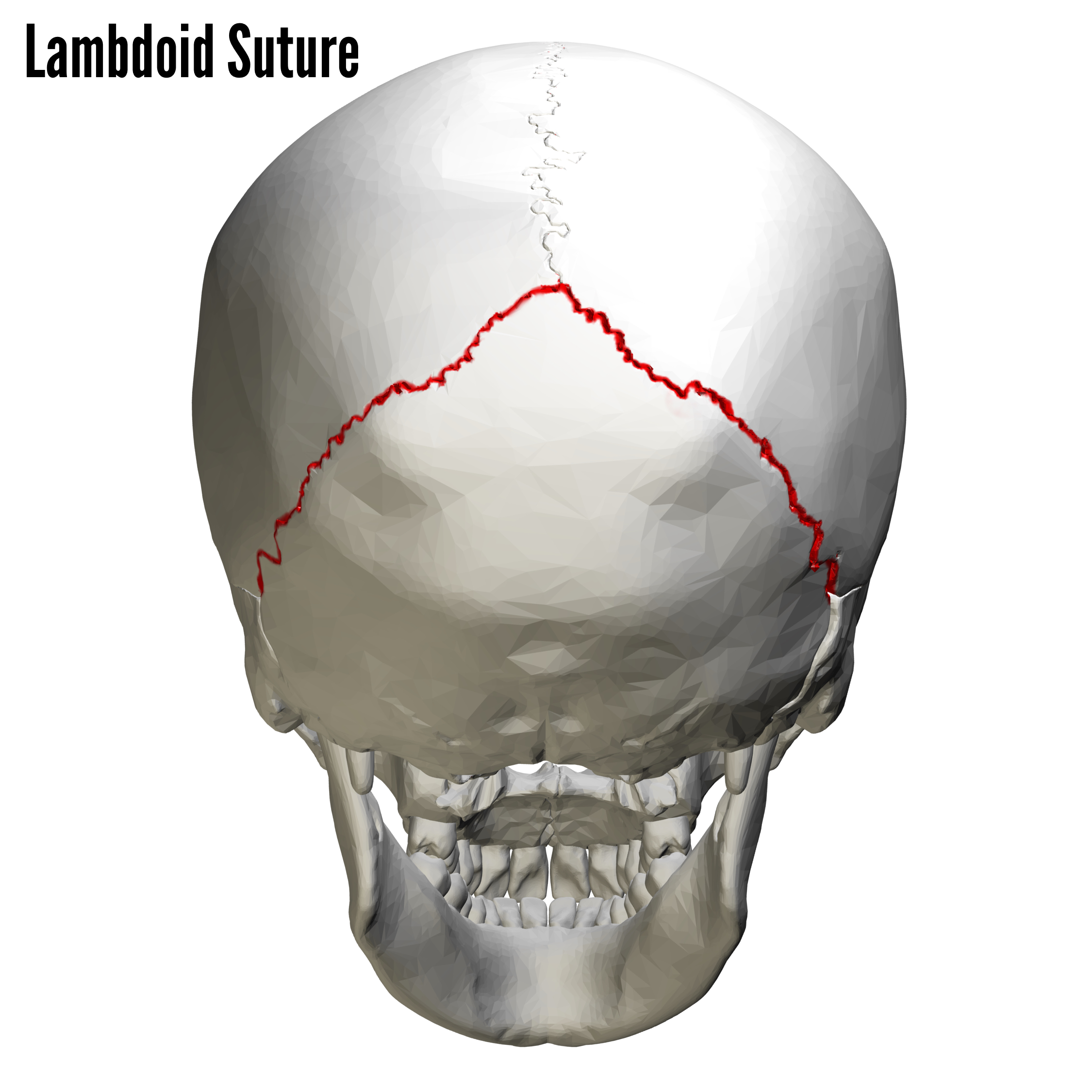
I Can Feel A Little Mass Protruding Out When I Touch The Back Of My Skull On Both Sides Of The Hemispheres Is This A Tumor Quora from qph.fs.quoracdn.net The frontal, parietal, temporal and occipital bones are joined at the cranial sutures. Skull bones aren't fused together at birth. In order to be light, the skull is made up by flat and irregular bones, and has hollow spaces called the sinuses. The bbc is not responsible for the content of external websites. Skull anatomy | with labels. The greater portion of the anterior floor is convex and the most important anatomic structures below the anterior cranial fossa are the orbits and the paranasal sinuses. It supports and protects the face and the brain. The skull is a bony structure that supports the face and forms a protective cavity for the brain.
Foramina inside the body of humans and other animals.
Skull anatomy and skull bones. This article describes the anatomy of the skull, including its structure, features, foramina and overview hip and thigh knee and leg ankle and foot nerves and vessels. The frontal, parietal, temporal and occipital bones are joined at the cranial sutures. Foramina of the skull and the structures that pass through. It is comprised of many bones, formed by intramembranous ossification, which are joined together by sutures (fibrous joints). Excluding ear ossicles, it is made of 22 bones. Anatomical structures of the skull include: The skull bones can be classified into two groups: So, the human skull consists of 23 bones. The skull has a single occipital condyle.7 the skull consists of five major bones: Learn about anatomy skull with free interactive flashcards. This anatomic region is complex and poses surgical challenges for otolaryngologists and neurosurgeons alike. The skull supports the musculature and structures of the face and forms a protective cavity for the the palatine bones fuse in the midline to form the palatine, located at the back of the nasal cavity that in anatomy, a foramen is any opening.
Related posts of bone of back of skull. Anatomical structures of the skull include: Looking at it from the inside it can be subdivided into. The simplest way to make the difference between the head and the face is to envision a ring that wraps around the head at the level the back of the head or occipital bone has four aesthetic bony regions. The skull is a bony structure that supports the face and forms a protective cavity for the brain.
Bones Of The Skull Skull Osteology Anatomy Geeky Medics from geekymedics.com The cranial vault denotes the top, sides, front, and back of the cranium. It offers protection to the brain, eye balls, inner ears, and nasal passages. Learn about anatomy skull with free interactive flashcards. Skull anatomy | with labels. The skull is the bony skeleton of the head. It supports and protects the face and the brain. The skull base is the inferior portion of the neurocranium. The temporal bone connects to the occipital bone in human skull from the front.
Learn about the anatomy of the skull bones and sutures as seen on ct images of the brain.
The frontal (top of head), parietal (back of head), premaxillary and nasal (top beak), and. The skull includes the upper jaw and the cranium. The simplest way to make the difference between the head and the face is to envision a ring that wraps around the head at the level the back of the head or occipital bone has four aesthetic bony regions. The base of the skull (or skull base) forms the floor of the cranial cavity and separates the brain from the structures of the neck and face. The skull begins to form prior to week 12 of embryogenesis. In order to be light, the skull is made up by flat and irregular bones, and has hollow spaces called the sinuses. Learn about anatomy skull with free interactive flashcards. The skull bones can be classified into two groups: Skull reshaping is done on any of the structures that lie above the face. The skull or known as the cranium in the medical world is a bone structure of the head. Anatomical structures of the skull include: The greater portion of the anterior floor is convex and the most important anatomic structures below the anterior cranial fossa are the orbits and the paranasal sinuses. The temporal bone connects to the occipital bone in human skull from the front.
It offers protection to the brain, eye balls, inner ears, and nasal passages. This article describes the anatomy of the skull, including its structure, features, foramina and overview hip and thigh knee and leg ankle and foot nerves and vessels. Foramina of the skull and the structures that pass through. The temporal bone connects to the occipital bone in human skull from the front. Human anatomy for muscle, reproductive, and skeleton.
External Aspect Of Base Of Skull from www.netterimages.com Human skull from the front. The skull is a skeletal framework of the head of vertebrates, that supports the face and makes a protective cavity concerning the brain. The bbc is not responsible for the content of external websites. Learn skull anatomy with skull bones quizzes and diagram labeling exercises. The skull begins to form prior to week 12 of embryogenesis. Learn about anatomy skull with free interactive flashcards. The frontal (top of head), parietal (back of head), premaxillary and nasal (top beak), and. Looking at it from the inside it can be subdivided into.
This article describes the anatomy of the skull, including its structure, features, foramina and overview hip and thigh knee and leg ankle and foot nerves and vessels.
The base of the skull (or skull base) forms the floor of the cranial cavity and separates the brain from the structures of the neck and face. Foramina of the skull and the structures that pass through. These joints fuse together in adulthood. Learn about the anatomy of the skull bones and sutures as seen on ct images of the brain. Excluding ear ossicles, it is made of 22 bones. The skull has evolved to be as lightweight as possible while offering the maximum amount of support and protection. Learn about anatomy skull with free interactive flashcards. The skull is the bony skeleton of the head. So, the human skull consists of 23 bones. The skull performs vital functions. Skull reshaping is done on any of the structures that lie above the face. Anatomical structures of the skull include: The frontal, parietal, temporal and occipital bones are joined at the cranial sutures.


0 Komentar